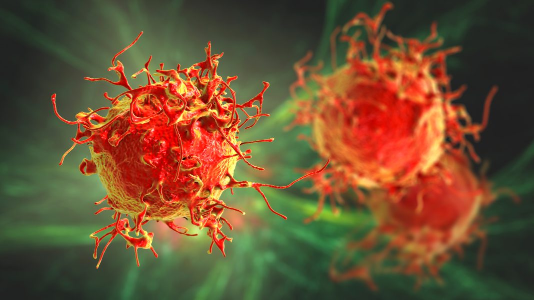
Scientists from the University of California (UC), San Diego, say they have discovered a process in which liver cells share molecules via vesicle exchange in order to multiply under conditions that would suppress cell proliferation. They also say they have discovered evidence that this process occurs in various types of cancer cells, which may lead to a new approach to tackling treatment resistance in cancer.
The findings are published in eLife, in an article titled, “Identification of CD133+ intercellsomes in intercellular communication to offset intracellular signal deficit.”
“CD133 (prominin 1) is widely viewed as a cancer stem cell marker in association with drug resistance and cancer recurrence,” wrote the researchers. “Herein, we report that with impaired RTK-Shp2-Ras-Erk signaling, heterogenous hepatocytes form clusters that manage to divide during mouse liver regeneration. These hepatocytes are characterized by upregulated CD133 while negative for other progenitor cell markers.”
“Understanding cell proliferation is a fundamental issue in both cancer research and biomedical science as a whole,” said Gen-Sheng Feng, PhD, a professor of pathology at UC San Diego School of Medicine and of molecular biology at UC San Diego School of Biological Sciences. “While we studied the liver here because of its incredible regenerative potential, we’ve also found these vesicles in other types of cancer cells. We believe that this could be a universal mechanism that drives resistance to cancer therapies that target cell proliferation.”
“Cells in the liver multiply more quickly and effectively than any other cells in the body, which makes the liver the ideal place to study the biological processes that control cell division,” said Feng. “These are the same processes that go awry in cancer, and so one promising approach to treating cancer is targeting cell proliferation.”
In previous research, Feng and his team observed that a minority of liver cells in mice could still proliferate even when the cells were genetically engineered to lack a critical signaling enzyme required for cell proliferation. This enzyme, called Shp2, helps liver cells know when it’s time to divide during liver regeneration.
By clustering together and exchanging tiny, protein-wrapped packages called vesicles, liver cells are able to communicate and share the biochemical materials needed to multiply, allowing them to proliferate even without Shp2.
“We’ve known for a long time that liver cells have other ways of regenerating even when lacking Shp2, and this study shows us how they do it,” said Feng.
The researchers also uncovered evidence that cancer cells may use the same strategy to resist treatment and continue to divide.
“We believe we’ve discovered an important strategy that tumors use to resist therapy,” said Feng. “This discovery could prove to be a powerful new target for cancer treatment, particularly as part of combination therapies to mitigate treatment resistance.”
The findings suggest a new way of thinking about cancer initiation, progression, and recurrence.
“Our findings could tell us that a cancer cell’s ‘stemness,’ or ability to initiate and reinitiate tumors, isn’t a central part of its identity, but a temporary state that can be induced or possibly even turned off,” said Feng. “This could be a brand-new way of looking at tumor recurrence that could open doors for new treatments.”


